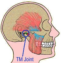
The temporomandibular joint (TMJ) is located just
in front of the ear. This joint is made up of the
lower jawbone (mandible) and the temporal bone of
the skull. The condyle of the mandible is that portion
which fits into the joint space. This condyle has
a disc composed of cartilage that sits on top of
the condyle. The disc prevents damaging bone-to-bone
contact during normal functioning of the TMJ. People
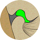
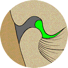
who suffer from a temporomandibular disorder (TMD)
may have this protective disc made up of cartilage
displaced off the condyle:
Normal Joint
Displaced Disc
In such cases, the TMD sufferer often has clicking,
popping, or grinding noises that can be heard during
normal functioning of the TMJ. People with these symptoms
should never go untreated as continual degeneration
of the TMJ will occur, possibly bringing debilitating
pain.
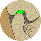
Persons suffering from TMD may also have a disorder
which differs from the displaced disc described above.
This type of disorder occurs when the condyle of the
mandible posteriorly positioned in the joint space:
Normally Positioned
Posteriorly Positioned
To understand why this creates the painful syndrome
of TMD, let us briefly explain the tissues that exist
in this joint space posterior to the condyle. Blood
vessels, nerves, and connective tissue are abundant
in this area. The external carotid artery, a large blood
vessel carrying blood from the heart to the brain, passes
through this area. The temporal-auricular nerve is one
of a number of nerves existing in this area as well.
When a condyle is posteriorly positioned in the TMJ,
it compresses and damages these tissues of nerves, blood
vessels, and connective tissue. Many persons with a
condyle, which is posteriorly compressed against these
delicate soft tissues, will develop frequent headaches.
With the passing of time, these headaches increase in
frequency and intensity.
This is why it is extremely important that all youngsters
with a deficient or retruded jaw be treated with the
functional appliances when they are young. These functional
orthodontic appliances will position the jaw so that
the TMJ’s are not damaged during normal functioning.
Early interceptive functional orthodontic treatment
of these cases helps create healthy temporomandibular
joints. Historically, treatment has often involved bicuspid
extraction or the use of cervical headgear. These classical
methods should rarely be used because of the likelihood
of trapping the mandible posteriorly, setting the stage
for painful disorders of the TMJ in later life.
Causes of TMJ
Lower jaw trapped back in a posterior position
An improper bite
Direct blow to the jaw or head
Whiplash injury from a rear end collision
Extraction of back teeth
Missing back teeth
Excessive clenching or grinding of the teeth
A whiplash injury, which occurs by a collision from
the rear, is one cause of TMJ disorders. Unique to this
injury is the absence of a direct blow to the head or
jaw. In this situation, the mouth opens excessively
wide as the head is snapped back. The joints of the
jaw dislocate if the collision from the rear is sufficiently
forceful. In addition, the soft tissues posterior to
the condyle (blood vessels, nerves, and connective tissue)
are compressed and damaged. Research by Arrington and
Garcia documented that 95% of their research subjects
suffered TMJ abnormalities after sustaining a whiplash
injury. In a similar study, Pressman found that 88%
suffered TMJ abnormalities.
Excessive clenching and grinding of the teeth when dealing
with tension and stress often cause TMD. This type of
TMJ disorder first begins in the jaw muscles. The clenching
and grinding tire the muscles and trigger spasms. This
produces pain and ultimately a TMD. Myofacial pain dysfunction
is the term used to describe this set of circumstances.
Because TMD mimics many other medical problems, the
TMD symptoms often go undiagnosed. If your physician
finds no underlying conditions for symptoms such as
frequent headaches, eye pain, or ear pain, he could
consider the possibility of a TMJ disorder. Your physician
can recommend that you consult a dentist who is properly
trained in diagnosing TMD and non-surgical treatment
of TMD. Surgical treatment is to be avoided as it builds
scar tissue in the TMJ. This scar tissue creates more
compression against the delicate soft tissues(blood
vessels, nerves, and connective tissue) posterior to
the condyle, which will in turn eventually increase
the intensity of the headaches and other symptoms of
TMD. The famed Mayo Clinic no longer allows surgical
treatment of TMD in their clinic because of this reason.
Symptoms of TMJ
Clicking or popping noises in the TMJ
Chronic Headaches
Ear pain or ringing in the ears
Jaw pain
Eye pain
Facial pain
Difficulty in chewing or opening the mouth
Jaws locking closed or locking open
Neck/shoulder pain
Misaligned teeth
Dizziness
It is estimated that more than 40 million Americans
suffer from one or more of these symptoms. Most sufferers
will not usually exhibit all these symptoms. Some persons
may not have symptoms severe enough to justify treatment.
However, about one of every eight Americans suffer frequent
headaches and pain severe enough to disrupt their normal
daily routine. If you suspect that you have a TMD and
need help, contact Fletcher Dental and TMJ Clinic. There
is hope for you!
Treatment of TMJ
TMD sufferers most severely damage their TMJ’s
while eating and sleeping. In order for healing to occur
in the TMJ’s, the condyles of the lower jaw must
not be allowed to compress the soft tissues posterior
to the condyles while eating or sleeping. Treatment
progresses through two phases.
Phase I treatment is accomplished through the
use of two different orthotic appliances. The daytime
appliance allows the TMD sufferer to eat without damaging
the tissues in the posterior joint space. The nighttime
appliance is designed so that the tissues in the posterior
joint space are not damaged during movements of the
jaw during sleep. These two appliances must be worn
24 hours a day for about nine months. These appliances
place the jaw and its condyles in a new pain-free position.
This nine-month period is necessary in order to allow
healing of the damaged tissues in the posterior TMJ
space.
Phase II treatment occludes the lower and upper
teeth together with the lower jaw in the new pain-free
position. Phase II treatment is best done by orthodontic
or prosthetic methods. If orthodontics is the chosen
phase II method, approximately 18 months treatment time
is needed. The treatment time for the prosthetic method
is much shorter. It can involve the placement of up
to 20 crowns on the upper and lower teeth. The goal
of phase II treatment is to stabilize the bite in the
new pain-free position so that the teeth, muscles, and
joints can work together without strain. Drs. James
and Matthew Fletcher have the training and experience
to provide phase II treatment using both the orthodontic
and the prosthetic method.
What should I do if I suspect TMD?
If you suspect that you suffer from TMD, call Fletcher
Dental and TMJ Clinic. There is hope for you. Drs. James
and Matthew Fletcher have received their training through
the American Association of Functional Orthodontics,
American Academy of Cranialfacial Pain, and the TMJ
Institute of America.